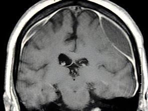
Chronic Subdural Hematoma
A Chronic Subdural Hematoma (CSDH) is a condition in which blood gradually collects between the outer covering of the brain (dura) and the surface of the brain. It usually develops over several weeks following bleeding from small blood vessels, often after a minor head injury. In some cases, patients may not remember any injury at all.
Chronic subdural hematoma is more common in older adults and may also occur in people taking blood-thinning medications or those with conditions that increase the risk of bleeding. Because the collection can gradually increase in size and put pressure on the brain, timely diagnosis and appropriate treatment are important.
Under the expert care of Dr Garga Basu, patients receive comprehensive, evidence-based evaluation and individualised treatment for chronic subdural hematoma, based on the size of the hematoma, symptoms, neurological condition, underlying risk factors, and overall health.
Causes and Risk Factors of Chronic Subdural Hematoma:
A chronic subdural hematoma can develop when small blood vessels between the brain and its outer covering are stretched or damaged, allowing blood to accumulate gradually.
Common causes and risk factors include:
- Minor or previously unnoticed head injuries
- Falls or accidents, particularly in older adults
- Use of blood-thinning or antiplatelet medicines
- Conditions that affect blood clotting
- Brain shrinkage associated with ageing, which can stretch small blood vessels
- Previous brain surgery or other neurological conditions in selected cases
- Increased risk of falls or repeated minor head trauma
In some patients, no clear history of head injury may be identified.
Symptoms of Chronic Subdural Hematoma:
Symptoms of chronic subdural hematoma may develop gradually and can sometimes be mistaken for normal ageing or other neurological conditions.
Symptoms may include:
- Persistent or worsening headache
- Difficulty walking or problems with balance
- Weakness or numbness, particularly on one side of the body
- Changes in speech
- Memory or concentration difficulties
- Confusion or changes in behaviour
- Drowsiness or increased sleepiness
- Vision problems
- Seizures
- Difficulty performing normal daily activities
- Loss of consciousness in severe cases
Symptoms may become progressively worse as the hematoma increases in size and places pressure on the brain. Sudden deterioration, seizures, loss of consciousness, or new weakness requires immediate medical attention.
Diagnosis of Chronic Subdural Hematoma:
Accurate diagnosis is essential to determine the size and location of the hematoma and assess the degree of pressure on the brain.
Depending on the patient’s condition, evaluation may include:
- Detailed medical and neurological assessment
- Review of any previous head injury or fall
- CT scan of the brain
- MRI of the brain in selected cases
- Blood tests to assess clotting and other relevant factors
- Review of medications, particularly blood-thinning or antiplatelet medicines
Brain imaging helps identify the location, size, and characteristics of the hematoma and assists in determining the most appropriate treatment.
Treatment for Chronic Subdural Hematoma:
Treatment depends on the size of the hematoma, severity of symptoms, neurological findings, rate of progression, and overall health of the patient.
Treatment may include:
- Close observation and follow-up with repeat brain imaging in selected patients with small or minimally symptomatic hematomas
- Management or adjustment of contributing medications when medically appropriate
- Correction of underlying blood-clotting abnormalities when required
- Surgical drainage: A procedure to remove the accumulated blood and reduce pressure on the brain in patients who require intervention
- Burr-hole drainage: A commonly used minimally invasive procedure in which one or more small openings are made in the skull to drain the hematoma
- Craniotomy: A larger surgical opening may be considered in selected patients, particularly when the hematoma is organised, recurrent, or not adequately treated with less invasive drainage
- Postoperative monitoring and follow-up to detect recurrence or other complications
Recovery and Follow-Up After Chronic Subdural Hematoma:
Recovery after treatment varies depending on the patient’s age, neurological condition, size and duration of the hematoma, and the presence of other medical conditions.
Many patients experience improvement in symptoms after successful drainage, although recovery may take time, particularly when the hematoma has caused prolonged pressure on the brain.
Some patients may require:
- Physiotherapy to improve strength, walking, and balance
- Occupational therapy to support daily activities
- Speech or cognitive rehabilitation when required
- Regular neurological follow-up
- Repeat CT or MRI scans to monitor recovery or detect recurrence
Why Choose Dr Garga Basu for Chronic Subdural Hematoma Care?
- Expertise in the diagnosis and management of neurological and neurosurgical conditions
- Patient-focused and evidence-based treatment planning
- Comprehensive neurological assessment and advanced brain imaging
- Individualised treatment based on the size, location, and clinical impact of the hematoma
- Appropriate use of minimally invasive and surgical treatment options when required
- Careful postoperative monitoring and follow-up
- Strong emphasis on patient safety, timely intervention, and long-term neurological outcomes
FAQs
1. Can a minor head injury cause a chronic subdural hematoma?
Yes. Even a minor head injury can sometimes cause bleeding from small blood vessels, particularly in older adults. Symptoms may develop gradually over weeks, and some patients may not remember having experienced an injury.
2. What are the warning signs of chronic subdural hematoma?
Warning signs may include persistent headache, difficulty walking, balance problems, weakness on one side, confusion, memory changes, speech difficulties, increasing drowsiness, seizures, or changes in behaviour. New or worsening neurological symptoms require prompt medical evaluation.
3. How is chronic subdural hematoma treated surgically?
A common treatment is burr-hole drainage, in which a small opening is made in the skull to drain the collected blood. Depending on the characteristics of the hematoma and the patient’s condition, other surgical approaches, including craniotomy, may be considered.
4. Can chronic subdural hematoma come back after surgery?
Yes. Recurrence can occur in some patients even after successful surgical drainage. Regular clinical follow-up and, when appropriate, repeat brain imaging can help detect recurrence early.
5. Who is at higher risk of developing a chronic subdural hematoma?
Older adults are at increased risk, particularly after falls or minor head injuries. The risk may also be higher in people taking blood-thinning or antiplatelet medicines and in individuals with conditions that affect blood clotting.
6. Can a chronic subdural hematoma cause weakness or difficulty walking?
Yes. Pressure from the hematoma can affect brain function and may cause weakness, difficulty walking, balance problems, or other neurological symptoms. Appropriate treatment may help improve these symptoms depending on the extent and duration of brain compression.