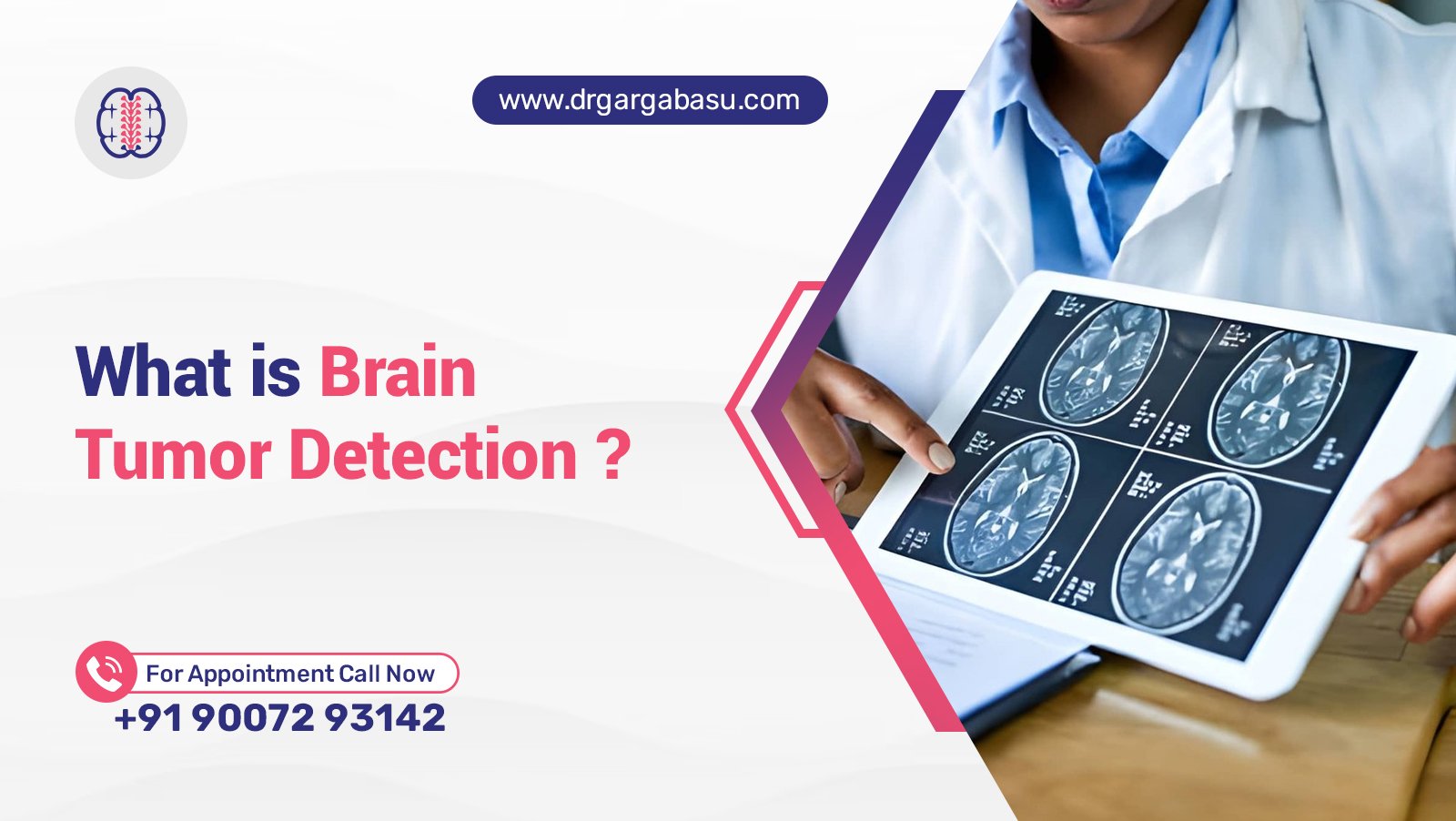
Brain tumor detection refers to the process of identifying abnormal cell growths within the brain using advanced medical technologies and expert clinical evaluation. Early detection of brain tumors dramatically improves treatment success, reduces neurological damage, and raises five-year survival rates. Integrating magnetic resonance imaging, computer‑aided diagnosis, and machine learning models equips health professionals with precise tools to spot lesions before symptoms worsen.
The human brain demands careful monitoring; subtle changes can signal malignancy that escalates quickly. When clinicians employ high‑resolution MRI scans, functional magnetic resonance imaging, and positron emission tomography, they capture detailed images of brain tissue that reveal tumor type and location. Combining these medical images with artificial intelligence accelerates tumor classification and supports accurate brain tumor diagnosis.
In this comprehensive guide we explore how detection methods work, why early diagnosis matters, common warning signs, diagnostic pathways, and current treatment options. Readers will gain a clear understanding of the role of technology, the spectrum of brain tumor types, and actionable steps for patients and caregivers.
Key Takeaways
- Early detection of brain tumors via MRI and AI‑driven analysis greatly enhances prognosis and expands treatment options.
- Warning signs such as persistent headache, seizure, and visual changes often indicate the need for immediate medical imaging.
- Advanced diagnostics including tumor classification, molecular profiling, and machine learning improve the accuracy of brain tumor diagnosis.
Understanding Brain Tumor Detection
Understanding brain tumor detection involves recognizing how modern techniques identify abnormal growths in the brain. Traditional methods rely on neurological examinations and symptom assessment, while contemporary approaches prioritize high‑resolution medical image acquisition.
Definition and Scope
Brain tumor detection encompasses the use of MRI scans, CT scan, and PET imaging to locate and characterize neoplasms within the brain. Detect brain tumors early prevents irreversible cognitive impairment and increases survival chances.
Role of Medical Imaging
Medical image analysis provides the foundation for tumor identification. MRI offers superior soft‑tissue contrast, revealing tumor borders and edema. CT scan supplies rapid assessment of bone involvement, and PET highlights metabolic activity to differentiate malignant from benign lesions.
Emerging AI and Machine Learning
Machine learning algorithms process thousands of images of brain, learning patterns that distinguish tumor types. Convolutional neural network models, such as AlexNet, automatically segment lesions, enabling rapid tumor classification and supporting radiologists in diagnosis.
Importance of Early Detection in Brain Tumor Management
Early detection in brain tumor care directly influences patient outcomes, treatment planning, and quality of life. Prompt identification allows clinicians to devise a personalized treatment plan before the tumor compromises vital functions.
Impact on Survival Rates
Studies show that patients diagnosed at an early stage experience a higher five‑year survival rate compared with those identified after symptom progression. Early detection of brain cancer reduces the risk of metastasis and limits malignant spread within the brain.
Influence on Treatment Options
When a tumor is identified early, less aggressive treatment options may suffice. Surgeons can perform minimally invasive stereotactic surgery, radiologists can apply focused radiation therapy, and oncologists can select targeted therapy or immunotherapy based on tumor genetics.
Reducing Neurological Deficits
Detecting brain tumors before they exert pressure on critical regions preserves cognition, speech, and motor control. Early intervention minimizes the need for extensive resection, thereby safeguarding the patient’s functional independence.
Common Warning Signs and Symptoms
Recognizing the early indications of brain tumor development enables timely medical evaluation. Both general and specific symptoms signal the need for immediate imaging.
Seven Warning Signs of Brain Cancer
- Persistent headache that worsens in the morning or with activity.
- Unexplained seizures or convulsions.
- Gradual loss of vision or new visual distortions.
- Difficulty speaking or understanding language.
- Weakness or numbness in limbs, especially on one side.
- Changes in personality, memory, or cognition.
- Unexplained nausea, vomiting, or balance problems.
Six Additional Signs of Brain Tumor
- Sudden hearing loss or ringing in the ears.
- Frequent dizziness or vertigo.
- Unexplained fatigue or lethargy.
- Hormonal imbalances causing irregular menstrual cycles.
- Unusual sensory perception such as tingling or phantom smells.
- Difficulty concentrating on routine tasks.
Diagnostic Pathway and Technologies
Diagnosing brain tumors follows a systematic approach that integrates clinical assessment, imaging, and pathology to achieve an accurate brain tumor diagnosis.
Imaging Modalities – MRI, CT, PET
Brain MRI provides detailed cross‑sectional images of brain tissue, enabling radiologists to locate lesions and assess tumor type. CT scans are valuable for detecting calcifications and bone involvement. Positron emission tomography using radioactive tracer highlights metabolic hotspots, assisting in differentiating aggressive tumors.
Biopsy and Molecular Testing
When imaging suggests malignancy, neurosurgeons perform a stereotactic brain biopsy to obtain tissue samples. Pathology examines cellular architecture, while molecular testing identifies biomarkers such as IDH mutation or MGMT promoter methylation, guiding targeted therapy decisions.
Tumor Classification and Type Identification
Tumor classification categorizes neoplasms into glioma, meningioma, schwannoma, and others. Identifying the tumor type influences prognosis and treatment. For example, glioblastoma represents a high‑grade malignant tumor, whereas a benign meningioma may require only observation.
Treatment Landscape and Prognosis
Effective treatment integrates surgery, radiation, chemotherapy, and emerging targeted therapies to address the diverse biology of brain tumors.
Surgery, Radiation Therapy, Chemotherapy
Complete surgical resection offers the best chance for cure in accessible tumors. Radiation therapy, including stereotactic radiosurgery, eliminates residual cells. Chemotherapy agents such as temozolomide complement radiation, especially for high‑grade gliomas.
Targeted Therapy and Immunotherapy
Targeted therapy exploits specific molecular abnormalities, while immunotherapy stimulates the patient’s immune system to attack tumor cells. Clinical trials evaluate the efficacy of checkpoint inhibitors and CAR‑T cells for recurrent brain cancer.
Rehabilitation and Quality of Life
Post‑treatment rehabilitation focuses on restoring motor function, speech, and cognitive abilities. Multidisciplinary care improves quality of life, reduces long‑term disability, and supports the patient’s return to daily activities.
Frequently Asked Questions
What is brain tumor detection?
Brain tumor detection involves using imaging studies, clinical assessment, and often AI‑based analysis to locate and characterize abnormal growths within the brain.
How to detect brain tumour early?
Early detection requires paying attention to warning signs, undergoing prompt MRI scans, and employing computer‑aided diagnosis tools that highlight subtle abnormalities.
What are the 7 warning signs of brain cancer?
The seven key signs include persistent headache, seizures, visual changes, speech difficulties, unilateral weakness, personality/cognitive shifts, and unexplained nausea or vomiting.
Can any treatments cure my brain tumor?
Cure potential depends on tumor type, grade, and location; complete surgical removal combined with radiation and chemotherapy offers the highest chance of long‑term remission for many tumors.
How many types of brain tumors?
There are dozens of brain tumor types, broadly categorized into gliomas, meningiomas, pituitary adenomas, schwannomas, and metastatic lesions, each with distinct behavior and treatment strategies.
Conclusion
Brain tumor detection and early diagnosis represent a critical frontier in neuro‑oncology. Leveraging high‑quality MRI, CT, PET, and machine learning‑driven tumor classification enables clinicians to identify malignancies at a stage where treatment options are most effective. Recognizing warning signs, undergoing timely imaging, and understanding the diagnostic pathway empower patients to seek care early, improve survival odds, and maintain quality of life. Continued advancements in AI, molecular profiling, and targeted therapies promise even greater precision in diagnosing and treating brain tumors, ultimately reducing the burden of this complex disease.