
Loading...

How Long for Trigeminal Neuralgia Medication to Take Effect and What to Expect

How Long for Trigeminal Neuralgia Medication to Take Effect and What to Expect

Trigeminal neuralgia involves intense, sudden facial pain caused by irritation of the trigeminal nerve. Managing this neurological disorder requires precise medication management and often surgical intervention to alleviate the paroxysmal attack cycles. Patients frequently experience electric-shock sensations across the cheek, jaw, or eye area.

Understanding the timeline for pain relief helps patients navigate their treatment for trigeminal neuralgia. Most medicine options aim to stabilize the nerve signals to prevent the characteristic stabbing sensations. While some drugs work quickly, others require gradual dose titration to reach therapeutic levels in the blood.

Key Takeaways

  • Medication Timeline: Most anticonvulsant drugs like carbamazepine take several days to weeks to reach full efficacy.
  • Primary Treatment: Carbamazepine remains the gold standard for initial trigeminal neuralgia treatment.
  • Surgical Options: If medicine fails, microvascular decompression or radiosurgery provide long-term relief.

How Long Does Trigeminal Neuralgia Medication Take to Work?

The time required for medicine to provide significant pain relief depends heavily on the specific drug type and the individual patient’s metabolism.

Most patients notice initial changes within a few days, but full stabilization of the trigeminal nerve activity takes longer. Because doctors must avoid rapid side effect onset, they often start with a low dose and increase it slowly. This process, known as titration, ensures the body tolerates the drug before it reaches the necessary concentration to block the abnormal pain signals.

Carbamazepine Efficacy Timelines

Carbamazepine typically requires several days to a few weeks to achieve optimal control over trigeminal neuralgia pain.

Patients starting carbamazepine often follow a specific schedule. During the first week, the drug builds up in the bloodstream. While some feel relief within 48 to 72 hours, the full therapeutic effect often emerges after two weeks of consistent use. The neurologist monitors the patient closely during this phase to manage any dizziness or nausea that might occur. If the paroxysmal attacks persist, the doctor might adjust the dose or suggest adding another medicine like oxcarbazepine or gabapentin.

Other Common Medications for Facial Pain

Different pharmacological options offer varying onset times for managing neuropathic pain associated with trigeminal neuralgia.

  • Oxcarbazepine: This anticonvulsant often shows a similar timeline to carbamazepine, providing relief within days to weeks.
  • Gabapentin: Often used for nerve pain, this drug requires a gradual increase in dose over several weeks to manage chronic pain effectively.
  • Baclofen: This muscle relaxant may be added to an anticonvulsant regimen, typically working within a week to assist in neurological stability.
  • Lamotrigine: Used in some cases, this requires a very slow titration to prevent the risk of a serious rash, such as Stevens–Johnson syndrome.
  • Pregabalin: This drug targets calcium channels to reduce pain signals and often shows effectiveness within one to two weeks.

Understanding the Cause of Trigeminal Neuralgia

The primary cause of trigeminal neuralgia involves physical compression of the nerve root by a neighboring blood vessel.

In many cases, an artery or vein presses against the trigeminal nerve near the brainstem. This constant pressure damages the myelin sheath, which acts as insulation for the nerve. When this insulation fails, the nerve becomes hypersensitive. This irritation causes the nerve to send incorrect, intense pain signals to the brain. Other causes include multiple sclerosis, where nerve damage occurs due to demyelination, or a tumor, such as a neoplasm, pressing on the nerve. Occasionally, an injury or dental procedure can damage the trigeminal nerve, leading to atypical trigeminal neuralgia.

The Role of Nerve Compression

Nerve compression triggers the electrical instability that leads to the characteristic facial pain.

When a blood vessel makes contact with the nerve, every pulse of blood creates micro-trauma. This trauma disrupts how the nerve transmits information. Instead of smooth signals, the nerve produces sudden, sharp bursts. This condition is often categorized under nerve compression syndrome. Neuroimaging, such as an MRI, helps a neurologist identify if a specific artery is the cause. Identifying the exact location of the compression helps determine if surgical treatment options like microvascular decompression (MVD) are appropriate.

Trigeminal Nerve Anatomy and Symptoms

The trigeminal nerve consists of three main branches that distribute sensation across the face.

  • Ophthalmic Branch: Provides sensation to the eye, forehead, and upper cheek.
  • Maxillary Branch: Carries sensations from the middle of the face and upper teeth.
  • Mandibular Branch: Controls sensation around the jaw and lower teeth.

Symptoms vary depending on which part of the trigeminal nerve is affected. Common signs include paroxysmal attacks of pain, facial numbness, or paresthesia. Some patients experience orofacial pain that feels like a toothache, often leading to unnecessary dental surgery. In some instances, patients report hypersensitivity to touch, where even a light breeze or washing the face triggers an attack.

Surgical Treatment Options for Trigeminal Neuralgia

Surgical procedures offer a way to permanently address the physical cause of the nerve irritation when medicine fails.

When drugs no longer provide sufficient pain relief, patients often consult a neurosurgeon. Surgical options range from highly invasive craniotomy to minimally invasive procedures. The choice depends on the patient’s age, overall health, and the specific cause of the neuralgia. While surgery carries risks, it offers the potential for long-term relief from the debilitating pain of trigeminal neuralgia.

Microvascular Decompression (MVD)

Microvascular decompression is a surgical procedure designed to relieve the pressure on the trigeminal nerve root.

During MVD, a surgeon performs a craniotomy to access the area where the nerve meets the brainstem. The surgeon identifies the offending blood vessel and places a small cushion, such as Teflon, between the vessel and the nerve. This effectively stops the compression. MVD is highly effective but involves a more invasive approach. Recovery requires careful management of the incision site and monitoring for potential complications like hearing loss or cerebrospinal fluid leaks.

Stereotactic Radiosurgery and Other Procedures

Non-invasive or minimally invasive techniques provide alternatives for patients who cannot undergo major surgery.

Stereotactic radiosurgery, often using the Gamma Knife, delivers a concentrated beam of radiation to the trigeminal nerve root. This radiation causes minor damage to the nerve, which eventually interrupts the transmission of pain signals. It is a common choice for elderly patients or those with multiple comorbidities. Other options include:

  • Glycerol Injection: A chemical injection that damages the nerve to reduce pain transmission.
  • Balloon Compression: A minimally invasive procedure using a balloon to compress the nerve root.
  • Radiofrequency Ablation: Uses heat to destroy part of the nerve.
  • Rhizotomy: A surgical method to sever the nerve fibers responsible for pain.

Managing Long-Term Pain and Expectations

Successful management of trigeminal neuralgia requires a multidisciplinary approach involving neurology and pain management specialists.

Living with chronic pain requires mental and physical adjustments. While medication controls the intensity of attacks, it rarely “cures” the underlying nerve damage. Many people with trigeminal neuralgia find that certain triggers, such as chewing, talking, or cold wind, exacerbate the condition. Keeping a diary of these triggers can help patients and doctors refine the treatment plan. In some cases, patients may experience facial numbness or weakness as a side effect of long-term drug use or surgical intervention.

Potential Side Effects of Medication

Most drugs used to treat trigeminal neuralgia carry specific side effects that require monitoring.

Because these medicines are anticonvulsants, they affect the central nervous system. Common side effects include:

  • Dizziness: Often occurs when starting or increasing the dose.
  • Nausea: Can be managed by taking medicine with food.
  • Ataxia: Coordination issues or clumsiness.
  • Somnolence: Increased sleepiness or fatigue.
  • Cognitive Changes: Difficulty concentrating or “brain fog.”

Frequently Asked Questions

How long does it take for carbamazepine to work for trigeminal neuralgia?

Carbamazepine typically begins to reduce pain within a few days, but achieving full therapeutic control usually takes one to two weeks of consistent use. Doctors often start with a low dose to mitigate side effects before gradually increasing it to the optimal dose for your specific needs.

Does trigeminal neuralgia go away with medication?

Medication often manages the symptoms effectively, but it does not always “cure” the underlying cause of the nerve irritation. For many, the medicine provides long-term relief, while for others, the effectiveness may wane over time, requiring a change in treatment options or surgical intervention.

How long does it take for the trigeminal nerve to repair itself?

The trigeminal nerve does not typically “repair” itself in the sense of reversing the damage caused by compression. Instead, treatments aim to either remove the source of irritation or stabilize the nerve’s electrical activity so it no longer sends incorrect pain signals to the brain.

How to permanently stop trigeminal neuralgia?

Permanent relief often requires addressing the physical source of the pain through surgical procedures. Microvascular decompression (MVD) is one of the most effective ways to stop the pain by physically separating the blood vessel from the nerve. Other permanent or long-term methods include stereotactic radiosurgery and various forms of nerve ablation.

Are there support groups for people with trigeminal neuralgia?

Yes, many patients find comfort and practical advice through various support groups. Online forums, patient advocacy organizations, and local neurological health care groups provide spaces to connect with others managing this specific type of facial pain and chronic pain disorder.

Conclusion

Navigating the complexities of trigeminal neuralgia requires patience and a proactive approach to medical diagnosis and treatment. While the initial period of starting carbamazepine or other medicines can be challenging due to titration and side effects, these drugs remain vital for managing the intense pain of trigeminal neuralgia. If pharmacological management fails to provide sufficient relief, surgical options like MVD or Gamma Knife radiosurgery offer significant hope for long-term stabilization. Always work closely with a neurologist or neurosurgeon to develop a personalized treatment plan that addresses your unique symptoms and improves your quality of life.
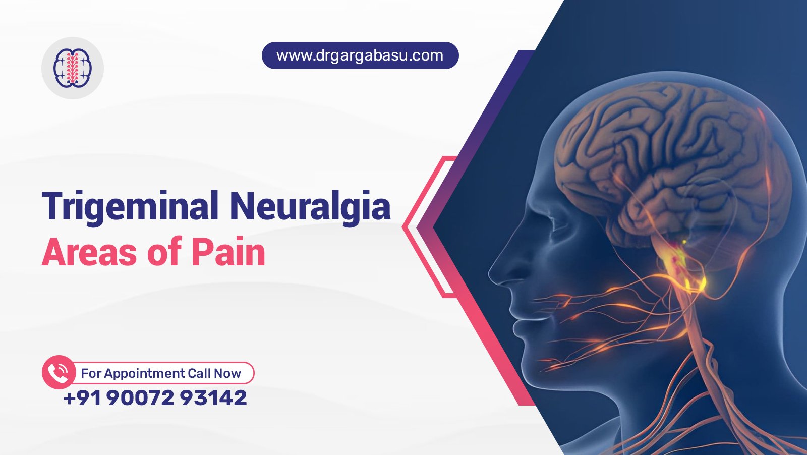

Trigeminal Neuralgia Areas of Pain and Their Significance in Diagnosis

Trigeminal neuralgia presents as brief, electric shock‑like attacks that strike one side of the face. The pattern of pain localization—forehead, cheek, jaw or palate—guides clinicians toward the correct diagnosis and influences treatment planning. Understanding which branch of the trigeminal nerve is affected helps differentiate classic TN1 from the atypical TN2 variant, identifies possible vascular compression, and alerts physicians to underlying conditions such as multiple sclerosis or neoplastic lesions.

Patients with trigeminal neuralgia often report that routine activities like brushing teeth, chewing, or even a gentle breeze trigger a paroxysmal attack. The sudden, intense facial pain can evolve into chronic pain if left untreated, impacting sleep, mood, and overall quality of life. Early recognition of pain distribution, combined with high‑resolution magnetic resonance imaging, streamlines the management of trigeminal neuralgia and reduces unnecessary procedures.

Key Takeaways

  • Precise pain mapping identifies the involved branch of the trigeminal nerve and directs appropriate imaging.
  • Vascular compression of the trigeminal nerve root remains the most common cause of classic trigeminal neuralgia, while demyelinating disease drives the idiopathic form.
  • Targeted interventions—microvascular decompression, radiosurgery, or pharmacologic therapy—depend on pain distribution, underlying pathology, and patient preference.

Distribution of Pain and Clinical Implications

Forehead and Peri‑Orbital Region (V1 Branch)

The ophthalmic division produces stabbing sensations on the forehead, scalp, and upper eyelid. Pain in this area often signals compression by the superior cerebellar artery or a cavernous sinus lesion. Neurological examination may reveal reduced corneal reflex, prompting urgent neuroimaging.

Cheek and Mid‑Facial Region (V2 Branch)

Maxillary involvement generates electric bursts in the cheek, upper lip, and upper gums. This distribution frequently accompanies arteriovenous malformation or a meningioma at the foramen rotundum. Patients with trigeminal neuralgia report that brushing or washing the face provokes attacks.

Jaw, Lower Teeth, and Chin (V3 Branch)

Mandibular pain targets the lower jaw, lower teeth, and anterior tongue. Compression by an aberrant artery near the mandibular foramen, or a tumor at the cerebellopontine angle, often produces V3‑dominant symptoms. Chewing, speaking, or yawning trigger intense paroxysmal episodes.

Pathophysiology and Etiologic Factors

Vascular Compression and Idiopathic Forms

In most cases, a pulsatile blood vessel exerts pressure on the trigeminal nerve root, generating ephaptic coupling between adjacent fibers. The resulting neuropathic pain manifests as classic trigeminal neuralgia. When imaging fails to reveal a compressive vessel, the condition is labeled idiopathic trigeminal neuralgia.

Multiple Sclerosis and Demyelination

Patients with multiple sclerosis experience demyelination of the trigeminal nerve within the brainstem. This demyelination disrupts normal conduction, creating a secondary form of trigeminal neuralgia that often presents at a younger age. The disease‑related variant may coexist with vascular compression, compounding symptom severity.

Neoplastic and Structural Causes

Neoplasms such as acoustic neuromas, meningiomas, or epidermoid cysts can compress the trigeminal nerve at the root entry zone. Structural anomalies, including a congenital bone spur or a cavernous sinus thrombosis, also produce focal pain. Detailed neuroimaging differentiates these etiologies from pure vascular compression.

Diagnostic Approach

Clinical Evaluation

A thorough history captures trigger patterns, pain intensity, and distribution. Physical examination assesses sensation across the three branches, corneal reflex, and motor function of the jaw. Patients with trigeminal neuralgia typically describe brief, shock‑like attacks lasting seconds, recurring dozens of times daily.

Neuroimaging Techniques

High‑resolution magnetic resonance imaging with constructive interference in steady state (CISS) sequences visualizes the trigeminal nerve root and surrounding blood vessels. Findings of a looped artery contacting the nerve confirm compressive etiology. Absence of visible compression supports an idiopathic diagnosis.

Electrophysiological Tests

Trigeminal somatosensory evoked potentials (TSEP) gauge conduction delays across the nerve. Prolonged latency suggests demyelination, while normal latency favors vascular compression. Electrophysiology supplements imaging when structural lesions remain equivocal.

FAQ

Is trigeminal neuralgia painful?

Trigeminal neuralgia causes intense, electric shock‑like facial pain that interrupts daily activities.

What are the hallmarks of trigeminal neuralgia?

The hallmarks include brief, recurrent attacks; triggers such as brushing, chewing, or wind; unilateral involvement of a single branch; and normal neurological exam between episodes.

How is trigeminal neuralgia diagnosed?

Diagnosis relies on a detailed symptom history, branch‑specific sensory testing, and high‑resolution magnetic resonance imaging to identify vascular compression or alternative lesions.

How common is trigeminal neuralgia?

Incidence estimates range from 4 to 13 per 100,000 individuals, with prevalence increasing after age 50.

Are there support groups for people with trigeminal neuralgia?

National neurological societies and online patient forums provide education, emotional support, and resources for patients with trigeminal neuralgia.

Therapeutic Options and Management Strategies

Pharmacologic Therapy

First‑line agents include carbamazepine, oxcarbazepine, and lamotrigine. Adjunctive drugs such as gabapentin, pregabalin, and baclofen reduce attack frequency.

Surgical Interventions

Microvascular decompression relocates the offending artery away from the trigeminal nerve root, offering long‑term relief. Alternative surgeries—balloon compression, radiofrequency rhizotomy, and stereotactic radiosurgery—target the nerve directly.

Radiosurgery Considerations

Evidence suggests that increasing the nerve length within the treatment volume enhances pain control, yet careful dosing prevents excessive facial numbness.

Conclusion

Mapping the precise area of facial pain remains essential for accurate diagnosis of trigeminal neuralgia. Distinguishing vascular compression from demyelinating or neoplastic causes directs appropriate management of trigeminal neuralgia, whether through medication, microvascular decompression, or radiosurgical techniques. Early identification of pain distribution, combined with advanced imaging, reduces the burden of chronic facial pain and improves quality of life for affected patients.

Can Trigeminal Neuralgia Be Bilateral and What It Means for Patients

Trigeminal neuralgia (TN) presents as sudden, electric‑shock‑like pain that strikes the face with alarming intensity. The condition commonly targets one side of the face, yet a subset of patients experiences pain on both sides, a pattern known as bilateral trigeminal neuralgia. Understanding the mechanisms, diagnostic challenges, and therapeutic pathways for bilateral involvement equips patients and clinicians to make informed decisions.

Neurology specialists emphasize that bilateral disease does not signal a different disorder, but rather a more complex expression of the same nerve compression syndrome. Emerging case series and surgical reports reveal that microvascular decompression (MVD) can achieve lasting pain relief even when both trigeminal nerves are affected. This article dissects the epidemiology, diagnostic criteria, and treatment options, while answering the most pressing patient questions.

Key Takeaways

  • Bilateral trigeminal neuralgia occurs in a minority of patients, often linked to vascular compression or multiple sclerosis.
  • Microvascular decompression remains the most reliable surgical intervention for permanent pain relief in both unilateral and bilateral cases.
  • Comprehensive management blends medication, minimally invasive procedures, and psychosocial support to sustain quality of life.

Understanding Bilateral Trigeminal Neuralgia

Bilaterality describes pain that affects the left and right sides of the face, either simultaneously or sequentially. Research estimates that 5‑10 % of patients with trigeminal neuralgia develop bilateral symptoms during the disease course. The underlying cause frequently involves vascular compression of the trigeminal nerve root entry zone by an artery such as the superior cerebellar artery.

pain that strikes the face

Pathophysiology of Bilateral Involvement

The trigeminal nerve conveys sensory information from the face to the brainstem. When a blood vessel contacts the nerve, pulsatile pressure creates demyelination, enabling ectopic electrical discharges. In bilateral cases, two separate vessels compress each nerve, or a single vascular loop extends across the midline. Multiple sclerosis can also produce demyelination within the trigeminal pathways, raising the likelihood of pain on both sides.

Primary Bilateral Trigeminal Neuralgia vs. Sequential Onset

Primary bilateral trigeminal neuralgia describes simultaneous pain on both sides at diagnosis. Sequential onset refers to a patient who first experiences unilateral pain, then later develops contralateral symptoms. A report of two cases highlighted successful bilateral microvascular decompression after sequential onset, underscoring the need for repeat imaging when new pain emerges.

Diagnosis and Classification of Trigeminal Neuralgia

Accurate diagnosis hinges on detailed patient history, neurological examination, and high‑resolution magnetic resonance imaging (MRI). The International Classification of Headache Disorders defines classic trigeminal neuralgia by paroxysmal attacks triggered by light touch, chewing, or speaking. Atypical trigeminal neuralgia includes constant background pain.

Imaging Strategies

High‑resolution MRI with constructive interference in steady state (CISS) sequences visualizes the relationship between arteries and the trigeminal nerve. Detection of vascular compression guides surgical planning, especially when bilateral involvement is suspected.

Differential Diagnosis

Neurologists differentiate trigeminal neuralgia from dental pain, temporomandibular joint dysfunction, cluster headache, and neuropathic conditions such as post‑herpetic neuralgia. A thorough physical examination rules out neoplasm, skull base lesions, and demyelinating disease. When MRI reveals plaques in the brainstem, multiple sclerosis becomes a probable contributing factor.

Treatment Options for Bilateral and Unilateral Cases

Management begins with pharmacologic therapy, proceeds to minimally invasive procedures, and culminates with surgical intervention if pain persists.

Medication Management

First‑line agents include carbamazepine and oxcarbazepine, both sodium channel blockers that diminish ectopic firing. Gabapentin and pregabalin serve as adjuncts when side effects limit dosage. Lamotrigine offers an alternative for patients intolerant to carbamazepine. Anticonvulsant therapy achieves pain relief in 70‑80 % of cases, yet long‑term use may provoke hyponatremia or dizziness.

Minimally Invasive Procedures

When medication fails, percutaneous techniques target the trigeminal root. Balloon compression, radiofrequency rhizotomy, and glycerol injection each create a controlled lesion that interrupts pain signals. Radiosurgery, such as Gamma Knife, delivers focused radiation to the nerve, producing gradual pain reduction over months.

Microvascular Decompression

Microvascular decompression directly addresses the compressive artery, inserting a Teflon pad to separate the vessel from the nerve. Bilateral MVD involves two separate craniotomies or a single extended exposure, depending on surgeon preference. Long‑term studies report permanent pain relief in 80‑90 % of patients, with low recurrence when the artery remains decompressed.

Living with Trigeminal Neuralgia

Effective pain management restores the ability to chew, speak, and engage socially without constant fear of an attack. Psychological support mitigates anxiety and depression that frequently accompany chronic facial pain. Support groups, both online and in‑person, provide peer‑to‑peer counsel, medication tips, and encouragement.

Quality of Life Considerations

Patients who achieve stable pain control report near‑normal daily activities, including work, exercise, and travel. Regular follow‑up with a neurology clinic monitors medication levels, side effects, and signs of recurrence.

Support Resources

National and regional organizations host monthly meetings, webinars, and forums dedicated to trigeminal neuralgia. Online communities share treatment experiences, surgical outcomes, and coping strategies, fostering a sense of belonging for individuals who often feel isolated.

Frequently Asked Questions

Is bilateral trigeminal neuralgia possible?

Yes, bilateral trigeminal neuralgia occurs in a minority of patients and may arise from dual vascular compression or demyelinating disease.

How to permanently stop trigeminal neuralgia?

Microvascular decompression offers the most reliable permanent solution, especially when imaging confirms vascular compression.

How often is trigeminal neuralgia bilateral?

Studies estimate bilateral involvement in 5‑10 % of all trigeminal neuralgia cases.

Can you have a normal life with trigeminal neuralgia?

Effective medication, minimally invasive procedures, or successful surgery enable many patients to resume normal activities and maintain employment.

Are there support groups for people with trigeminal neuralgia?

Numerous support groups exist online and in major medical centers, providing education, emotional aid, and advocacy.

Conclusion

Bilaterality adds complexity to trigeminal neuralgia but does not preclude durable relief. Accurate diagnosis, careful evaluation of vascular anatomy, and a stepped treatment algorithm—including microvascular decompression—equip patients to achieve lasting pain control. Ongoing research into nerve‑protective agents and refined surgical techniques promises further improvements in outcomes for both unilateral and bilateral presentations.

Trigeminal Neuralgia vs Postherpetic Neuralgia Key Differences Explained

Trigeminal neuralgia and postherpetic neuralgia represent two distinct neuropathic pain conditions that significantly impair quality of life. Trigeminal neuralgia originates from dysfunction of the trigeminal nerve, the largest cranial nerve responsible for facial sensation and motor functions. Postherpetic neuralgia arises as a complication of herpes zoster (shingles), a reactivation of the varicella zoster virus that causes chickenpox. Both conditions produce severe pain, but their underlying pathophysiology, clinical features, and treatment approaches differ markedly.

Accurate diagnosis is essential because mismanagement leads to prolonged suffering and inadequate pain relief. Trigeminal neuralgia typically presents with sudden, shock-like facial pain triggered by routine activities such as chewing or light touch. Postherpetic neuralgia manifests as persistent burning or stabbing pain in a dermatomal distribution after the acute herpes zoster rash resolves. Understanding these differences helps health professionals select appropriate pharmacotherapy, nerve blocks, or surgical interventions. This article provides a comprehensive comparison of trigeminal neuralgia and postherpetic neuralgia, covering pathophysiology, key symptoms, risk factors, and evidence-based treatment options.

  • Cause and origin differ – Trigeminal neuralgia stems from neurovascular compression of the trigeminal nerve root or demyelinating lesions, whereas postherpetic neuralgia results from nerve damage caused by varicella zoster virus reactivation in the dorsal root ganglion.
  • Pain quality and triggers vary – Trigeminal neuralgia produces paroxysmal, lancinating electric shocks triggered by light touch or movement; postherpetic neuralgia causes constant burning, aching, or stabbing pain with allodynia and hyperalgesia.
  • Treatment strategies diverge – First-line therapy for trigeminal neuralgia includes carbamazepine or oxcarbazepine and microvascular decompression for refractory cases; postherpetic neuralgia management relies on topical lidocaine, gabapentinoids, tricyclic antidepressants, and antiviral drugs during the acute phase.

Understanding Trigeminal Neuralgia

Trigeminal neuralgia is a unilateral orofacial neuropathic pain condition affecting one or more branches of the trigeminal nerve. The disorder involves the trigeminal nerve root and its distribution, often due to compression by a blood vessel at the root entry zone near the brainstem. Less commonly, multiple sclerosis or a tumor causes secondary trigeminal neuralgia. The incidence is approximately 4–13 per 100,000 people annually, with higher frequency in women and individuals over 50 years. Pain episodes are severe, brief, and recurrent, leading to major depressive disorder, anxiety, and insomnia.

Pathophysiology and Key Features

The hallmark of trigeminal neuralgia is sudden, severe, electric-shock pain limited to the distribution of the trigeminal nerve. The pain is paroxysmal and lancinating, lasting seconds to two minutes. Common triggers include light touch, chewing, talking, brushing teeth, or cold wind. Sensory loss is typically absent in classic trigeminal neuralgia, although some patients report mild hypoesthesia or altered thermal sensation. The condition can affect the ophthalmic (V1), maxillary (V2), or mandibular (V3) branches, with V2 and V3 most frequently involved. Diagnosis relies on clinical history and neurological examination; magnetic resonance imaging helps exclude secondary causes such as a lesion or demyelinating disease.

Types of Trigeminal Neuralgia: TN1 vs TN2

Trigeminal neuralgia is classified into two main types. TN1 (classic) is characterized by intense, stabbing, intermittent pain. TN2 (atypical) presents with constant aching, burning, or throbbing pain of lower intensity but longer duration. When comparing which is worse, TN1 episodes produce more severe acute pain, while TN2 causes persistent pain that disrupts daily function and sleep. Many patients with TN2 also experience superimposed paroxysms. Both types negatively affect quality of life, but TN1 is often considered more debilitating due to the unpredictable, excruciating nature of attacks. Treatment selection depends on the predominant pain pattern.

Understanding Postherpetic Neuralgia

Postherpetic neuralgia (PHN) is the most common complication of herpes zoster, defined as pain persisting for at least 90 days after the acute rash resolves. Approximately 10–20% of shingles patients develop PHN, with risk increasing with age and immunodeficiency. The varicella zoster virus remains latent in sensory neurons of the dorsal root ganglion; reactivation causes inflammation, blister formation, and nerve damage. PHN involves the peripheral nervous system and manifests as neuropathic pain in the affected dermatome, most often the thorax. The condition significantly impairs physical and psychosocial well-being, contributing to fatigue, weight loss, and mood disorders.

Causes and Risk Factors

The primary cause of postherpetic neuralgia is nerve injury from varicella zoster virus replication. After primary chickenpox infection, the virus enters a state of latency in the dorsal root ganglion or trigeminal ganglion. Reactivation, triggered by declining immunity from age, stress, or immunosuppression, leads to acute herpes zoster. During the acute phase, viral spread causes neuritis, hemorrhage, and necrosis of sensory neurons and supporting cells. Risk factors for PHN include age over 60, severe acute rash, prodromal pain, ophthalmic involvement (herpes zoster ophthalmicus), and inadequate antiviral therapy. Vaccination with the zoster vaccine reduces the incidence of shingles and PHN.

Symptoms and Diagnosis

The pain of postherpetic neuralgia is often described as burning, aching, stabbing, or like an electric shock. Allodynia (pain from a nonpainful stimulus) and hyperalgesia (increased sensitivity to a painful stimulus) are common. Patients may also report hypoesthesia, itch, or altered thermal sensation in the affected dermatome. The rash typically heals within two to four weeks, but dysesthesia persists. Diagnosis is clinical, based on a history of herpes zoster with residual pain. Neurological examination may reveal sensory loss or altered reflexes in the dermatome. No specific laboratory test confirms PHN; the diagnosis relies on temporal association with acute herpes zoster.

Key Differences Between Trigeminal Neuralgia and Postherpetic Neuralgia

Although both conditions fall under the category of neuropathic pain, their etiology, location, pain characteristics, and treatment differ substantially. The following comparison outlines the main distinctions for health professionals and patients.

  • Cause – Trigeminal neuralgia is usually caused by neurovascular compression of the trigeminal nerve root; postherpetic neuralgia results from viral nerve damage after herpes zoster.
  • Location – Trigeminal neuralgia is strictly unilateral along branches of the trigeminal nerve; PHN occurs in any dermatome, most commonly the thoracic region.
  • Pain quality – TN pain is paroxysmal, lancinating, and electric-shock–like; PHN pain is constant burning, aching, or stabbing with prominent allodynia.
  • Triggers – TN attacks are triggered by light touch, chewing, or talking; PHN pain is often continuous without specific triggers, though contact may worsen allodynia.
  • Sensory findings – TN typically has no sensory loss; PHN often involves hypoesthesia, hyperalgesia, and thermal sensation changes.
  • Treatment – TN first-line: carbamazepine, oxcarbazepine, or microvascular decompression; PHN first-line: topical lidocaine, gabapentinoids, tricyclic antidepressants, and antiviral drugs in acute phase.

Connection Between Shingles and Trigeminal Neuralgia

Shingles can affect the trigeminal nerve, leading to a condition called trigeminal postherpetic neuralgia. Herpes zoster involving the ophthalmic branch (herpes zoster ophthalmicus) accounts for 10–15% of cases and carries a high risk of PHN. In this scenario, the varicella zoster virus reactivates in the trigeminal ganglion, causing rash and pain in the distribution of the trigeminal nerve. While classic trigeminal neuralgia is not caused by a virus, the herpetic form mimics its symptoms. Magnetic resonance imaging may reveal pontine trigeminal T2-hyperintensity, suggesting herpetic etiology of trigeminal neuralgia. This finding helps differentiate viral-induced neuralgia from neurovascular compression. Antiviral therapy during acute herpes zoster reduces the risk of chronic pain.

Advanced Treatment Options

Botulinum Toxin Type A for Intractable Trigeminal Neuralgia

Botulinum toxin type A has emerged as a potential treatment option for intractable trigeminal neuralgia. Studies show that local injection into trigger zones or along the affected nerve branch reduces pain frequency and intensity. The mechanism involves blockade of nociceptive neurotransmitter release and reduction of peripheral sensitization. Although not a first-line therapy, botulinum toxin provides pain relief for patients unresponsive to carbamazepine or who cannot undergo microvascular decompression. More randomized controlled trials are needed to confirm efficacy and optimal dosing.

Acupuncture for Postherpetic Neuralgia

Acupuncture is considered an adjunctive treatment for postherpetic neuralgia. Systematic reviews suggest that acupuncture reduces pain intensity and improves quality of life when combined with standard pharmacotherapy. The therapy may modulate somatosensory pathways, promote endogenous opioid release, and reduce inflammation. However, evidence is limited by small sample sizes and variable methodology. Acupuncture is not a replacement for antiviral drugs or topical medications, but it can be a safe complementary option for patients with refractory pain or those seeking nonpharmacologic pain management.

Frequently Asked Questions

What is the difference between post-herpetic neuralgia and trigeminal neuralgia?

Postherpetic neuralgia is a chronic neuropathic pain condition caused by nerve damage from the varicella zoster virus after a shingles infection, typically affecting a single dermatome on the thorax or face. Trigeminal neuralgia is a facial pain disorder resulting from compression or irritation of the trigeminal nerve, producing brief, electric-shock attacks. The primary difference lies in etiology: viral versus mechanical, and in pain pattern: constant burning versus paroxysmal stabbing.