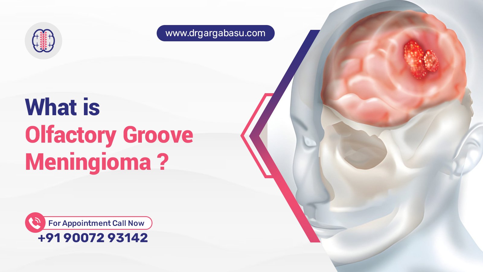
What Is Olfactory Groove Meningioma and How Is It Treated
Olfactory groove meningioma is a benign tumor that arises from the meninges, the protective layers surrounding the brain. The lesion grows in the anterior cranial fossa, near the cribriform plate and the olfactory nerve, often compressing the frontal lobes and causing loss of smell, headaches, and visual changes.
Because the tumor sits at the junction of the frontal bone and the cranial base, it can affect the optic nerve, the optic chiasm, and the surrounding arachnoid membrane. Early detection through MRI or CT scan imaging improves the chance of a successful resection and reduces the risk of recurrence. This article reviews the epidemiology, clinical presentation, diagnostic work‑up, and current treatment strategies for this rare brain tumor.
Key Takeaways
- Olfactory groove meningioma is a benign meningioma that commonly presents with anosmia, headache, and visual disturbance.
- High‑resolution MRI and CT imaging guide surgical planning, often involving a craniotomy or endoscopic approach.
- Combined treatment—complete surgical resection plus selective radiation therapy—offers the best long‑term control and survival.
Epidemiology and Risk Factors
How common is olfactory groove meningioma?
Olfactory groove meningioma represents 5–10 % of all intracranial meningioma cases, translating to less than 1 % of all brain tumors. Women develop the lesion more frequently than men, and incidence rises after the fifth decade of life.
Can smoking cause brain tumors?
Epidemiological studies show a modest association between long‑term tobacco exposure and increased risk of certain cancer types, including aggressive brain tumor subtypes. However, a direct causal link between smoking and olfactory groove meningioma remains unproven.
Prevalence of brain tumors in Connecticut
The Connecticut Cancer Registry reports an annual incidence of approximately 22 cases per 100 000 residents for all primary brain tumor diagnoses, aligning with national averages. Olfactory groove meningioma accounts for a small fraction of this figure.
Clinical Presentation and Diagnosis
Signs and symptoms
- Anosmia or reduced sense of smell.
- Frontal headache that worsens with Valsalva maneuvers.
- Visual field defects due to compression of the optic nerve or chiasm.
- Memory loss and mood changes from frontal lobe involvement.
- Seizures in up to 30 % of patients, reflecting cortical irritation.
Imaging and diagnostic work‑up
Contrast‑enhanced MRI provides the gold‑standard view, revealing a well‑defined, dural‑based tumor with a “tail” sign extending to the meninx. CT scan adds detail about hyperostosis of the frontal bone and calcifications. Advanced magnetic resonance angiography assesses vascular involvement, while biopsy remains rare due to the classic imaging appearance.
Pathology
Microscopic analysis shows meningothelial cells arranged in whorls, confirming a benign meningioma. Immunohistochemistry demonstrates EMA positivity and low Ki‑67 index, supporting the non‑malignant nature of the lesion.
Treatment Strategies
Surgical resection
Complete surgical removal remains the cornerstone of therapy. A craniotomy through a frontal or supra‑orbital keyhole approach provides direct access to the anterior fossa. In selected cases, a minimally invasive endoscopic technique reduces morbidity while allowing resection of the mass and involved dura. The goal is to remove the tumor with clear margins, preserving the optic nerve and olfactory tract when feasible.
Radiation therapy
Adjuvant radiation therapy is recommended for subtotal resection, atypical histology, or recurrent disease. Fractionated stereotactic radiotherapy delivers precise doses to the residual tumor while sparing surrounding brain tissue. In select patients, radiosurgery offers a non‑invasive alternative when surgery is contraindicated.
Can a meningioma be treated without surgery?
Observation with serial imaging is appropriate for small, asymptomatic meningioma lesions. Hormonal manipulation and targeted therapies are under investigation, but current evidence supports surgery as the primary curative option for symptomatic olfactory groove meningioma.
Management of recurrence
Recurrence rates range from 5–15 % after gross‑total resection. Repeat surgery, stereotactic radiosurgery, or combined modalities address regrowth. Long‑term follow‑up with annual MRI monitors for new lesions.
Prognosis and Survival
What is the survival rate for olfactory groove tumor?
Five‑year overall survival exceeds 90 % for patients undergoing complete resection of a benign meningioma. Functional outcomes depend on pre‑operative deficits; preservation of the optic nerve and olfactory function improves quality of life.
Factors influencing outcome
Age, tumor size, extent of resection, and histological grade dictate prognosis. Adjuvant radiation therapy improves control in high‑risk cases, while advanced age and tumor invasion of the cavernous sinus predict lower survival.
Supporting Patients and Caregivers
Helpful ways to support a loved one with a brain tumor
- Provide consistent emotional presence and active listening.
- Assist with appointment logistics, medication management, and symptom tracking.
- Encourage participation in support groups and neuro‑psychology services.
- Facilitate a balanced diet, adequate hydration, and rest to reduce fatigue.
- Advocate for clear communication between the patient and the multidisciplinary care team.
Frequently Asked Questions
How do you treat olfactory meningiomas?
Primary treatment involves surgical resection via a craniotomy or endoscopic approach, followed by selective radiation therapy for residual or recurrent disease.
Can brain tumors cause seizures?
Yes, cortical irritation from a brain tumor frequently triggers seizures, especially when the lesion involves the frontal or temporal lobes.
Can a meningioma be treated without surgery?
Observation with periodic imaging is viable for small, asymptomatic lesions; however, symptomatic meningioma requires operative intervention for definitive control.
How common is olfactory groove meningioma?
Olfactory groove meningioma accounts for 5–10 % of all intracranial meningioma cases, making it a rare but clinically significant tumor.
What are the different types of brain cancer?
Brain cancer classifications include astrocytoma, oligodendroglioma, glioblastoma, medulloblastoma, ependymoma, and meningioma, each with distinct histology and behavior.
Conclusion
Olfactory groove meningioma is a distinct benign tumor of the meninges that presents with olfactory loss, headache, and visual deficits. Robust MRI and CT imaging facilitate accurate diagnosis, while surgical resection—often via a craniotomy—offers the best chance for cure. Adjunctive radiation therapy enhances control in cases of subtotal removal or recurrence. Early intervention, multidisciplinary care, and comprehensive support for patients and caregivers collectively improve survival and quality of life for those affected by this unique brain tumor.